Introduction
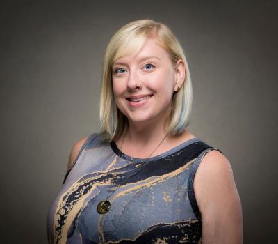
In a field where surgical outcomes are often unpredictable and chronic pain can persist despite multiple interventions, Morgan Giers is helping to reshape the landscape of spine care. As an assistant professor of bioengineering at Oregon State University and co-founder of the startup Spine by Design, Inc., Giers is leveraging predictive modeling, machine learning, and advanced imaging to bring precision and personalization to spinal treatment.
Her research is rooted in a simple but powerful question: Can we match the right patient to the right procedure — and do it before the first incision is made?
The problem with spine surgery
Spine surgery in the U.S. has a troubling track record. Procedures like spinal fusion and microdiscectomy often fail to relieve pain, with some patients undergoing multiple surgeries over numerous years—a population Giers refers to as “frequent flyers.” These patients bounce between specialists, often without lasting relief.
“The average age for spine surgery is in the early forties,” Giers said. “These are people who still want to run marathons or lift their kids. When surgery fails, it’s not just disappointing — it’s life-altering.”
Predicting reherniation — and preventing it
One of Giers’ most impactful projects to date centers on reherniation after microdiscectomy, a common surgery involving the removal of the portion of a herniated disc compressing a nerve root. Her team analyzed data from over 350 patients, identifying key risk factors such as disc height index, body mass index, lumbar lordosis angle, herniation type, and smoking status. Using a regression model, they predicted with 97% accuracy which microdiscectomy surgeries resulted in reherniation.
assistant professor of bioengineering
Blue Primary, Yellow Secondary
Furthermore, in a prospective study, Giers’ model helped reduce reherniation rates from 4% to 1%. But the results weren’t consistent across institutions — highlighting a deeper issue in spine care: measurement variability.
“Different people draw lines differently on spine images,” Giers said. “Even with the same instructions, the results vary. That’s a huge problem when you’re trying to build predictive models.”
To solve this, her lab, led by master’s student Sonia Ahrens, developed new measurement methods using the center-of-mass and orientation of discs, a more robust and error-tolerant method than previous clinical standards relying on discrete disc edges. These tools improved consistency and laid the groundwork for integration into neural networks for automated analysis.
From lab to startup: The birth of Spine by Design
This work emerged from Giers’ postdoctoral collaboration with surgeons at the Barrow Neurological Institute in Arizona, who had amassed large datasets but lacked the tools to analyze them. Initially brought in to run stats on the data for a journal article, Giers realized the potential for something bigger.
“I saw that the reherniation risk was predictable,” she said. “That’s when I knew we had something.”
Unfortunately, after starting at Oregon State, Giers repeated the study with multiple institutional partners and found the results were not repeatable because of the large variation between the way individual clinicians take spine measurements. That’s when she knew she could leverage her image-processing background to standardize spine measurements between institutes, not just as a precursor for the reherniation risk prediction, but for the entire spectrum of spine care.
Thus, the idea for Spine by Design was born.
Encouraged by Charla Triplett, who earned a master’s in bioengineering at Oregon State, has sat on the board of the School of Chemical, Biological, and Ecological Engineering’s Industry Advisory Board for 19 years, and is currently the CEO of Spine by Design, Giers transitioned from academic publishing to entrepreneurship. The company’s intellectual property centers on how spine measurements are taken, rather than the predictions themselves. This distinction allows Spine by Design to focus on standardizing imaging metrics, a critical step toward broad functionality and clinical adoption.
Today, the company is in its early stages. But its vision is bold: to integrate predictive modeling into hospital systems and electronic medical records, helping physicians make data-informed decisions.
Having already developed their alpha software in collaboration with Portland-based Synaptiq, the team’s next step is planning a multi-institutional trial to validate their models across diverse patient populations and imaging systems. The company has received grants from the National Science Foundation and Oregon State’s Advantage Accelerator, and is preparing for its first formal fundraise.
Phenotyping and machine learning
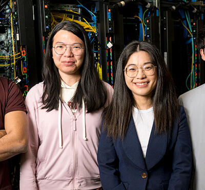
Another frontier in Giers’ research is phenotyping degenerative disc disease. By analyzing 21 variables from 95 pre-surgical patients, her team, led by former postdoctoral researcher Liudmila Bardonova and master’s student Joseph Chen, identified three distinct clusters with different tissue characteristics, demographics, and outcomes.
This work could revolutionize treatment selection, allowing physicians to tailor interventions based on a patient’s unique pathology rather than generalized symptoms.
“We treat all spine patients the same,” Giers said. “But not all back pain is the same. We need to understand where the degeneration starts and how it progresses to know the best course of treatment.”
A vision for intelligent spine care
Ultimately, Giers envisions a future where spine care is intelligent, personalized, and predictive. Patients would be evaluated using standardized metrics, matched to the best procedure, and monitored with AI-driven tools.
“We’re many years away,” she said. “But that’s my career goal, building a comprehensive system that guides each patient to their best possible outcome.”
Sidebar: Cryopreservation and the future of spine research

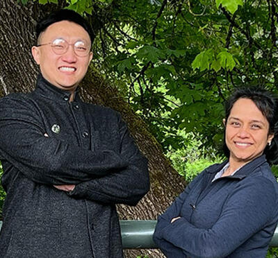
In collaboration with Adam Higgins, associate professor of bioengineering, Giers’ lab is working to improve the way that scientists study spinal disc degeneration — a leading cause of disability — by developing a method to preserve donated human intervertebral discs for research.
Picking up work initiated by Ward Shalash, PhD bioengineering ’22, bioengineering PhD student Rachel Thompson is now leading the way in applying a technique called cryopreservation, which involves freezing discs to -135°C to bring the cells into a suspended state. A major challenge is that without proper preparation, the chemicals used in the cryopreservation process can damage cells. To overcome this problem, Thompson applies compression to the discs before freezing, allowing the protective chemicals to penetrate more effectively—much like how a sponge soaks up liquid.
The results have been striking. Discs that are both compressed and cryopreserved show significantly higher cell viability compared with those that are frozen without compression. This breakthrough could dramatically improve the reliability of spine research and accelerate the development of new treatments for chronic back pain.