Introduction
Brian Bay had always been fascinated by life sciences but decided to pursue another passion, mechanical engineering, as an undergraduate at the University of California, Davis.
More than three decades later, in the serendipitous way that research and careers often take shape, Bay’s interests in machinery and medicine — and photography — have coalesced to push the boundaries of understanding for osteoarthritis.
His research has provided the first complete, cellular-level look at what’s going on in joints afflicted by a debilitating and costly disease that affects nearly one-quarter of adults in the United States.
“It happened the way most of the interesting stuff happens — a variety of experiences coming together in some unexpected way, a synthesis process more than a targeted endeavor,” said Bay, associate professor of mechanical engineering in the College of Engineering at Oregon State University. “If there’s one message that I hope alumni hear, it’s that supporting science and research environments — more than specific, targeted outcomes — is where most innovation comes from.”
When Bay was a graduate student — his bachelor’s, master’s, and doctoral degrees were all earned at UC Davis — he was offered funding to help with a research project on bone graft materials.
“The experience demonstrated to me that, among many other things, humans are highly sophisticated mechanical contraptions — but imperfect ones,” Bay said. “I stayed with orthopedic research, and my role was providing the engineering perspective, which is critical when the fundamental issue is mechanical breakdown.”
As a student, Bay focused on stress analysis, specifically through a computer-based method known as finite element modeling.
“It is a natural fit for biomechanics applications, because it’s suited to the complex geometries that bones and joints exhibit,” he said. “But it bothered me that published models were so simplistic compared to the real thing, and most of the models had no experimental validation.”
Digging deeper, Bay found publications on a nascent technique called digital image correlation that relied on photographic images of study samples subjected to loading.
“I was intrigued by the possibility of doing science by taking pictures of things, as I have always liked photography,” he said. “I began experimenting with bone samples, wrote basic correlation software, and found success in measuring tissue strains. When early research using X-ray tomography began to appear, I made an important connection: Volumetric strain measurement could be based on a 3D version of image correlation code.”
Bay described his discovery as digital volume correlation. The first demonstration that it worked came on bone tissue from a knee joint, and it’s now used to measure volumetric strain on many different types of materials.
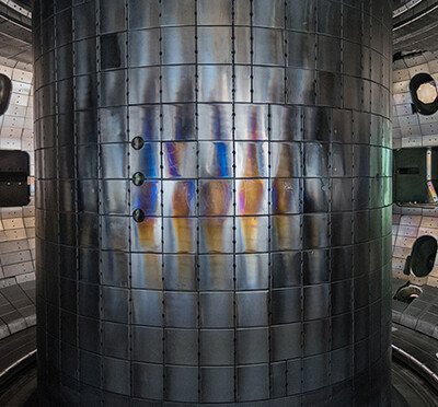
Using synchrotron X-ray tomography, Brian Bay and collaborators provide the first cellular-level functional evaluation of mouse bones and joints under mechanical loading. The study is paving the way to better understanding and treatment of arthritis.
“I was in a lab where I worked directly with clinicians who knew the patient and treatment side of the problem,” he said. “I also was working directly with actual tissue samples and got to see what they were really like. I also knew a bit about photography, was intrigued by X-ray tomography, had a background in mechanics, and could program a computer. These things all rolled together within an environment where trying new things was encouraged and supported.”
Bay’s most recent study, published in November 2019 in Nature Biomedical Engineering, opens the door to better understanding how interventions such as diet, drugs, and exercise affect a joint’s cells, which is important because cells do the work of developing, maintaining, and repairing tissue.
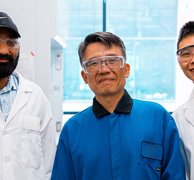
Bay and scientists from the Royal Veterinary College in London and University College London developed a sophisticated scanning technique to view the loaded joints of arthritic and healthy mice.
“Imaging techniques for quantifying changes in arthritic joints had been constrained by a number of factors,” he said. “Restrictions on sample size and the length of scanning time are two of them, and the level of radiation used in some of the techniques ultimately damages or destroys the samples being scanned. Nanoscale resolution of intact, loaded joints had been considered unattainable.”
Bay and collaborators including scientists from 3Dmagination Ltd. (UK), Edinburgh Napier University, the University of Manchester, the Research Complex at Harwell, and Diamond Light Source developed a way to conduct nanoscale imaging of complete bones and whole joints under precisely controlled loads.
To do that, they had to enhance resolution without compromising the field of view, reduce total radiation exposure to preserve tissue mechanics, and prevent movement during scanning.
“With low-dose pink-beam synchrotron X-ray tomography, and mechanical loading with nanometric precision, we could simultaneously measure the structural organization and functional response of the tissues,” Bay said. “That means we can look at joints from the tissue layers down to the cellular level, with a large field of view and high resolution, without having to cut out samples.”
Two features of the study make it particularly helpful in advancing the study of osteoarthritis, he said.
“Using intact bones and joints means all of the functional aspects of the complex tissue layering are preserved,” Bay said. “And the small size of the mouse bones leads to imaging that is on the scale of the cells that develop, maintain, and repair the tissues.
“It turns out the bet on using image data for measurement work was very fortunate as imaging technologies have continually improved and measurement capabilities have improved along with them.”
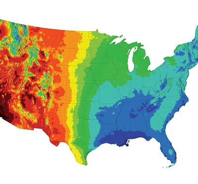
Osteoarthritis, the degeneration of joints, affects more than 50 million American adults, according to the Centers for Disease Control and Prevention. Women are affected at nearly a 25% rate, while 18% of men suffer from osteoarthritis.
As baby boomers continue to swell the ranks of the U.S. senior population, the prevalence of arthritis will likely increase in the coming decades, according to the CDC.
The CDC forecasts that by 2040 there will be 78 million arthritis patients, more than one-quarter of the projected total adult population; two-thirds of those with arthritis are expected to be women. Also by 2040, more than 34 million adults in the U.S. will have activity limitations due to arthritis.
“Osteoarthritis will affect most of us during our lifetimes, many to the point where a knee joint or hip joint requires replacement with a costly and difficult surgery after enduring years of disability and pain,” Bay said. “Damage to the cartilage surfaces is associated with failure of the joint, but that damage only becomes obvious very late in the disease process, and cartilage is just the outermost layer in a complex assembly of tissues that lie deep below the surface.”
Those deep tissue layers are where early changes occur as osteoarthritis develops, he said, but their basic biomechanical function and the significance of the changes are not well understood.
“That has greatly hampered knowing the basic disease process and the evaluation of potential therapies to interrupt the long, uncomfortable path to joint replacement,” said Bay, who saw his mother and grandmother undergo those types of procedures.
“This study for the first time connects measures of tissue mechanics and the arrangement of the tissues themselves at the cellular level — a significant advance, as methods for interrupting the osteoarthritis process will likely involve controlling cellular activity. It’s a breakthrough in linking the clinical problem of joint failure with the most basic biological mechanisms involved in maintaining joint health.”
The Engineering and Physical Sciences Research Council, Arthritis Research UK, the Medical Research Council, and the Diamond-Manchester Branchline at Diamond Light Source supported this research.